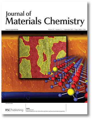
 Artwork for Journal of Materials Chemistry
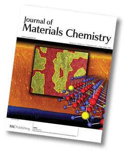
In late Summer 2012 I got the oppurtunity to do design work combined with my science work - I was invited to submit a suggestion for front page artwork for the scientific Journal "Journal of Materials Chemistry", published by the Royal society of chemistry. The end result, as it was printed on the inside front cover of the issue where me and co-authors published our findings on new materials based on aluminium, germanium and nitrogen in a paper (J. Mater. Chem. v22, nr33, p16761, 2012). According to the artwork instructions, the cover "needn't be scientifically sound", which gives some additional design freedoms such as colouring :) Below a step-by-step view of how the artwork was created is given. Most of the work was done in Photoshop.
More detailed scientific and technical description is given in separate paragraphs using smaller text...
The basis for the design was an scanning electron microscopy (SEM) image of a sample, which will be used as backround
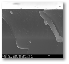
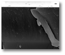
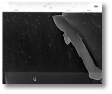
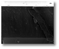
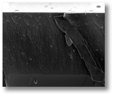
Field of view approximately 3 µm wide, i.e. roughly a 1/33 of the width of a human hair. Image on top is as it was saved from the instrument, and the following ones are different steps of image enhancement whereof only the first one would be permissable for normal scientific publications as the following ones are applied on selected areas.
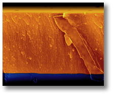
To make things somewhat prettier the image is thereafter colorised, with yellow/orange for the coating material and blue for the substrate on which the coating was grown. A dark fade was alos added top and bottom to create a border for the artwork.
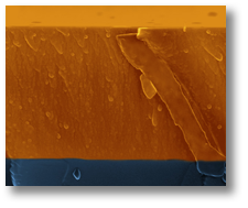
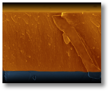
The second element of the design was a transmission electron microscopy (TEM) image, also enhanced and colourised:
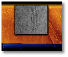
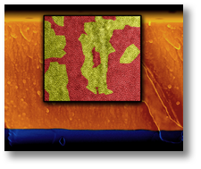
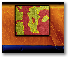
The TEM image has much higher resolution than the SEM imgage, it's field of view that is approximately 35 nm, i.e. 1000 times smaller than the SEM image in the background. The coluring shows the two different phases in the material: a crystalline particle phase (AlN) in yellow and an amorphous matrix phase (a-GeNx in red; as well as different grains in the crystalline phase - boundaries are marked with thick yellow lines. TEM micropscopy done by Magdalena Parlinska-Wojtan
The third and last element of the design was a atomic structure model made created using two programs: Diamond and POV-ray. The attained image was then tweaked with reagards to colour, but more importantly a tranparency fade was added to reinforce the feeling of an infinite crystal.
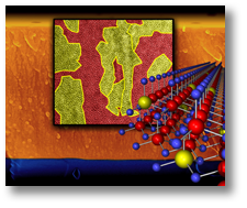
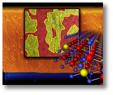
The structure model shows the structure of the crystalline AlN-phase. This model was created first using the program Diamond, and contains just short of 1000 atoms. The image was created in the ray-trace program POV-Ray. Bond lengths between the atoms are in the order of 2 Å, i.e. about 175 times shorter than the width of the TEM-image. The structure is in principle a wurzite AlN structure (Al red, N blue) with Ge atoms (yellow) in solid solution. The Final .PSD-file contained 26 layers, whereof 21 visible / active, and was about 260 MB large...
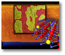
The image above right was sent to the publishers, and was then subsequently selected for the inside cover (J. Mater. Chem. v22, nr33, p16660, 2012) of issue in question, which then looks like this:
|